Case study
Case study Nero
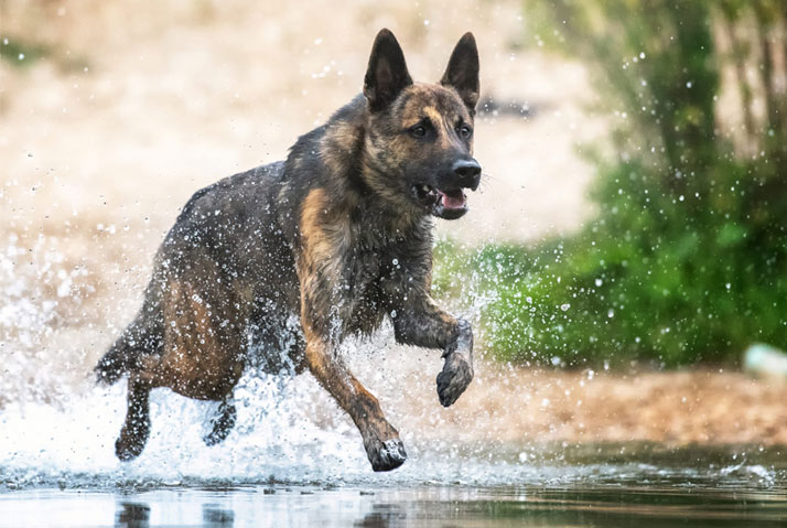
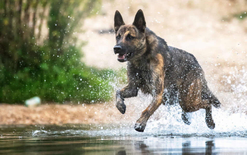
Dutch shepherd, male, 2 years old, has been lame on the left front for 12 weeks.
This real-life case is presented by the World Veterinary Organisation for Kinematically Controlled Orthopaedics, Neurology and Rehabilitation. Its founder and chairman of the board is Dr. med. vet. Patrick Blättler Monnier.
Initial problem
After the initial veterinary therapy, which had been initiated with the suspicion of a back problem, had not been able to cure the condition, Nero’s subsequent referral as an emergency at a clinic led to the assumption of a potential elbow pathology. However, this statement had not been substantiated with X-rays and the cause of the lameness, which until then had remained unclear, could only be revealed by the functional orthopaedic examination in Dr. Blättler Monnier’s practice.
Orthopaedic findings
Thus, during a functional orthopedic examination, Nero was found to have moderate muscle atrophy in the supraspinatus muscle on the left, as well as a limitation of extension and flexion of the left shoulder. In addition, the elbow joint showed mild heating, while ROM in the elbow – especially pro- and supination – was decreased, the back was stiff, and the thoracic and lumbar spine were hypomobile.
Image diagnostics
Subsequent sonography of the biceps tendon clearly showed that its bursa was very filled, so Nero was sedated for a CT study of the shoulder limbs to be sure. This clearly excluded a proc. Coronoideus (elbow dysplasia).

Therapy with subsequent sonography
Therapeutically, a tenotomy of the biceps tendon was performed and after six weeks a control sonography was initiated, which revealed a severed tendon but also the healing of the bursitis.
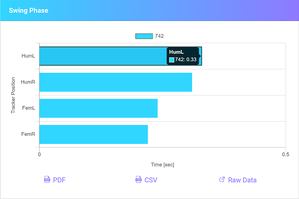
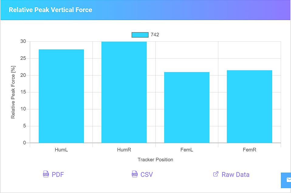
Functional movement measurement with LupoGait®
The functional movement measurement with the 4Dvets-system showed a reduction in the stance phase of the left shoulder limb, but a slight prolongation of the slope phase and an equally slight increase in the vertical acceleration (PVF).
These findings of the stance and swing phases as well as the PVF represent a clear sign of relief and are also considered evidence that the restoration of functionality of the left shoulder has not yet been fully achieved.

In the video sequence no lameness is seen and the movement presents as normal; however, on closer inspection it is noticeable that the left shoulder limb is not fully space-gripping and the loading is reduced compared to the right shoulder limb.
Follow-up therapy monitored by LupoGait®
The therapeutic solution could be a physiotherapeutic treatment as well as ACS-therapy with the autologous conditioned serum, the success of which can be monitored before and after the injection in an evidence-based manner with the the LupoGait®-system.