Case study
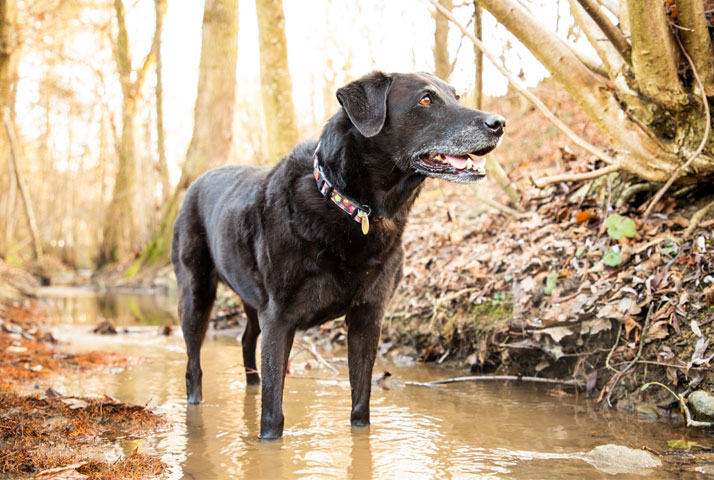
Case Study Jools
Labrador, female, spayed, 13 years old; was evaluated as healthy at the official HD/ED x-ray.
This real-life case is presented by the World Veterinary Organisation for Kinematically Controlled Orthopaedics, Neurology and Rehabilitation. Its founder and chairman of the board is Dr. med. vet. Patrick Blättler Monnier.
Initial problem
After an initial suspicion of a bone tumour, an unclear diagnosis and pain therapy with no results, the dog was first referred to Dr. Patrick Blättler Monnier’s canine practice for an orthopaedic examination in 2011 due to pass gait. There, Jools underwent mechanical orthopaedic manual release and the sessions were repeated in 2013 and 2015 before the first symptoms of overuse in the back became apparent in 2016. From then on, the back was also treated at annual intervals in conjunction with physiotherapy measures. In 2017, Dr. Blättler Monnier suspected elbow and toe arthrosis and was able to identify the problem of lameness, which recorded an increase at the beginning of 2020 and was localised on the right front.
Orthopaedic findings
The functional orthopedic examination revealed a severe regression of the shoulder muscles (supra- and infraspinatus) and a marked change in the elbows. In addition, the right shoulder also showed a restriction: especially the extension of the leg showed a considerable reduction.
Image diagnostics
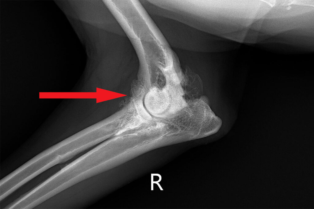
The subsequent X-rays of the shoulder girdle taken in the standing position revealed bilateral high-grade elbow and toe joint arthrosis as well as degenerative changes in the spine.
Functional movement measurement with LupoGait®
The kinematic examination confirmed the diagnosis by determining the stride and gait length, the extension and flexion of the limbs as well as their full mobility (range of motion).
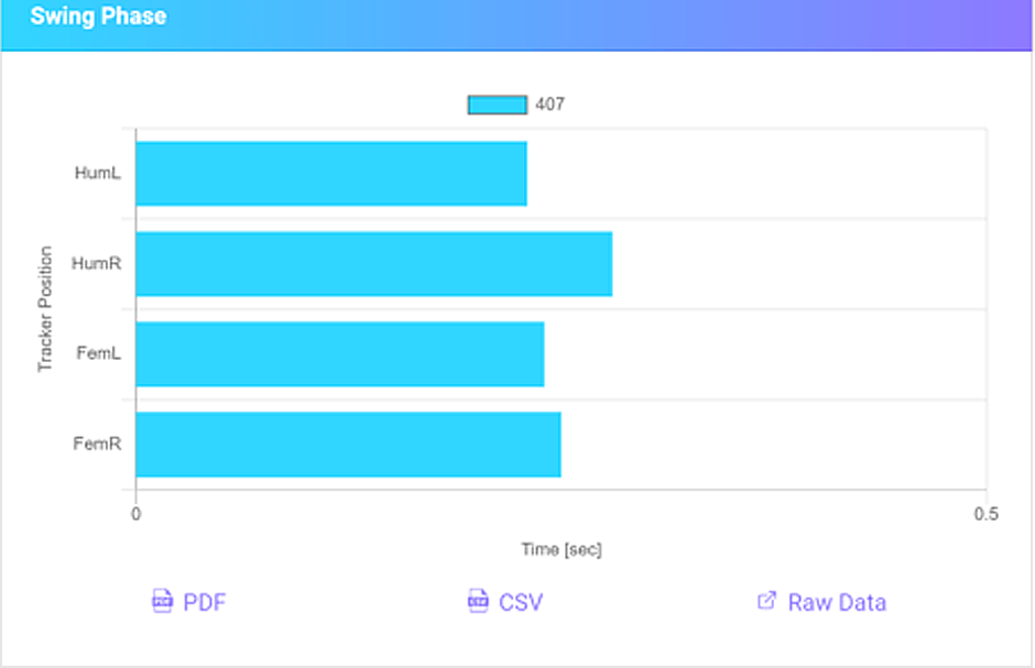
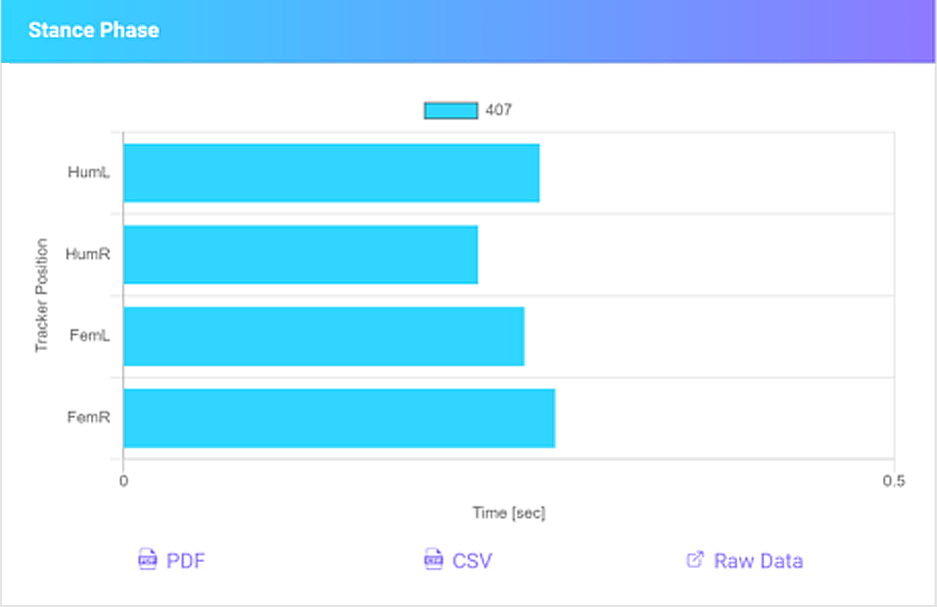
This showed that Jools was taking up eighty percent of the weight on the forehand, but only twenty percent on the backhand.
The straightening alone, followed by a control measurement, had again led to her being able to move freely and without strain and to the weight distribution being balanced (see video).
As can be seen from the X-ray, the overloading of Jool’s hindquarters was caused by a spondylosis as well as a clear defect of the intervertebral disc on the lumbar vertebra L7, which at the same time affected the exits of the nerve tracts. The dog instinctively tried to get pain relief by shifting her weight forward. The compensatory absorption of additional weight by the shoulder limbs led to edge pressure and increasing friction and triggered an inflammatory process in the joint.
Follow-up therapy monitored by LupoGait®
Thanks to the combination of X-rays and the evidence-based, objective movement analyses, the consequences could be clearly proven, and the subsequent targeted therapy could be monitored and successfully completed.