
A street dog fights hip dysplasia and pain
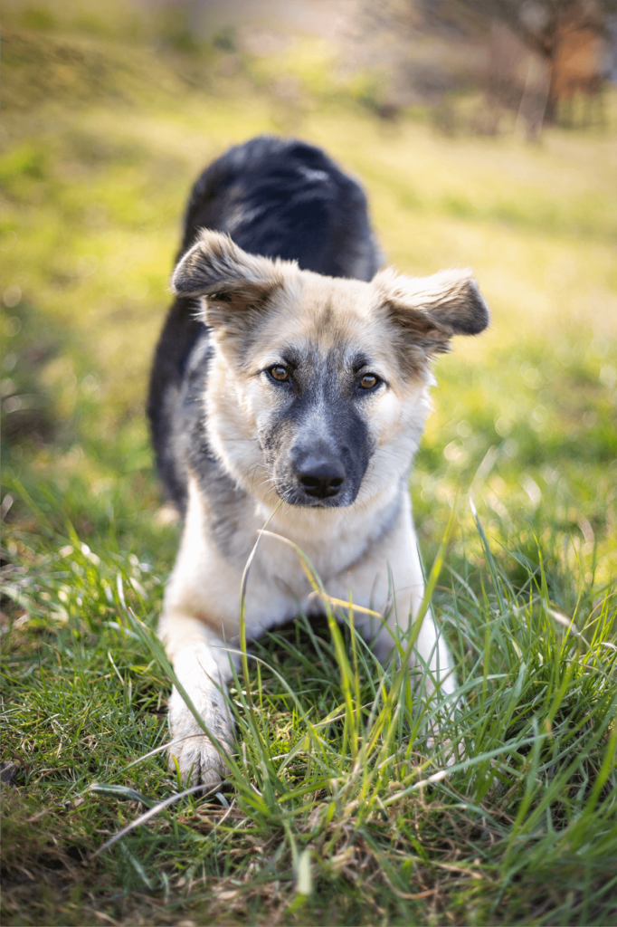
Frida’s journey begins as a street dog in Romania. As a young puppy, she was found in a rubbish bag. An international animal welfare organisation rescued her and brought her to Germany to her new family. Her new-found happiness lasted only a short time, however, because soon after her arrival she developed difficulties walking. Shortly after, she was no longer able to stand.
Her condition was diagnosed as bilateral hip dysplasia;
her condyle didn’t sit properly in the socket, often popping out. The cartilages rubbed against each other, which after a while destroys their natural shock-absorbing function and can lead to hip arthritis. All this caused Frida severe pain. A juvenile pubic symphysiodesis, a minimally invasive procedure on the growth plate, was to serve as a temporary solution. Frida was still too young to have an artificial hip joint.
Diagnosis and symphysiodesis
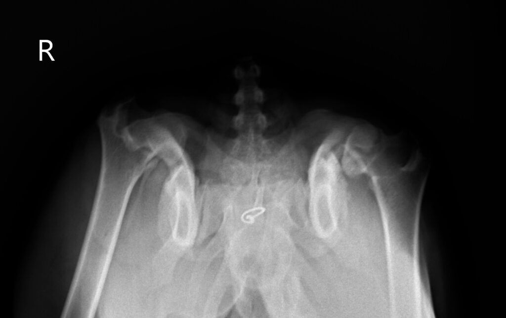
It is easy to see that the hip does not move in the socket in an extended position. Also in a frog position the hip is loose on both sides.
The third picture shows a distraction technique. The hip is spread and the looseness is tested at the same time. In Frida’s case the looseness is 1, which indicates a very loose hip.
Diagnosis and outlook
As a result of the diagnosis, we discussed the clinical picture and the resulting options with the owner. The owner immediately decided against euthanasia.
She was clearly informed that the further treatment would have to be divided into two steps. On the one hand, a short-term therapy, the symphysiodesis, to support Frida’s growth in the next months, to reduce her pain and to allow her a more normal socialisation.
In the medium term, two artificial hip joints will have to be inserted afterwards.
Furthermore, the owner was shown the exact costs and therapeutic consequences for the dog, as well as the duration and type of rehabilitation and regeneration.
Symphysiodesis – Frida’s first operation
Following the examinations, Frida immediately underwent a juvenile pubic symphysiodesis. This means that the pubic symphysis is surgically closed at an early stage. The technique itself is a relatively new procedure that was first successfully applied to guinea pigs and has also been successfully applied to canine patients for some years.
It is a prophylactic preventative surgery that is used primarily in growing dogs that show a great tendency to hip dysplasia and loose hips.
Laser surgery is used to cause a portion of the pubic symphysis to ossify. As a result, the lower, rear part no longer grows, while the upper part continues to form. This results in a better roofing of the femur bones than would have been the case without therapeutic measures – the stability of the joints is increased.
The symphysiodesis was successful, as was the post-operative check: Frida no longer walks with a severe limp.
Outlook
However, in Frida’s case, this preventive treatment is not enough to completely eliminate her HD. Despite the success of the surgery, it only provides a means to allow Frida to grow normally without pain. She should be able to move normally, at least with only a little lameness, for the next few months until she is fully grown. However, without major surgery, Frida’s hip will never be able to function like a normal one.
At a later date, when all the growth plates are closed, Frida’s treatment will continue.
Frida’s custom-made TEP – the left hip joint
At a later stage, when all the growth plates are closed, Frida’s treatment will continue.
The initial situation
Frida is now in Dr. Blättler-Monnier’s practice. It is early autumn 2022, 7 months after her successful symphysiodesis. She is again showing pain and lameness, especially on the back left side, after lying down and moving around, but also after playing with other dogs.
New hip joint from 3D printing
The reactive hip is the cause of Frida’s problems. Frida is now older – so an artificial hip joint for the left side was considered. The orthopaedic examination only confirmed this. However, no simple, cementless TEP (total endoprosthesis) was to be inserted. A 3D-printed TEP was created based on the bone and pelvic structure.
For this purpose, Frida was first referred for a CT study. The data collected was processed and used to create a model corresponding to the hip prosthesis. The model was tested to see if it could be attached to the bone and work correctly from a biomechanical point of view. The tests were successful – a prosthesis for the left side was created, which was inserted before the end of autumn 2022. Physiotherapeutic measures following the operation should help her to walk evenly.
Frida’s fight against hip problems continues: second operation necessary
Frida came through the operation well overall, and the control of the newly implanted hip was also successful. In January of this year, Frida again developed difficulties when walking: she again showed an irregular gait; in addition, she has pain in her right hip.
The left leg turns outwards and Frida is also sensitive at the back left, at the operated hip. The owner came for an orthopaedic re-evaluation.
In movement, the left, operated side turns outwards, while the right side shows a clear slope leg lameness. The overall movement is uneven as well as irregular.
Reactive hip and uneven leg length:
The challenges of Frida’s orthopaedic examination
The examination of the left pelvic limb, hip and knee are unremarkable and not painful. The right side, however, is quite reactive:
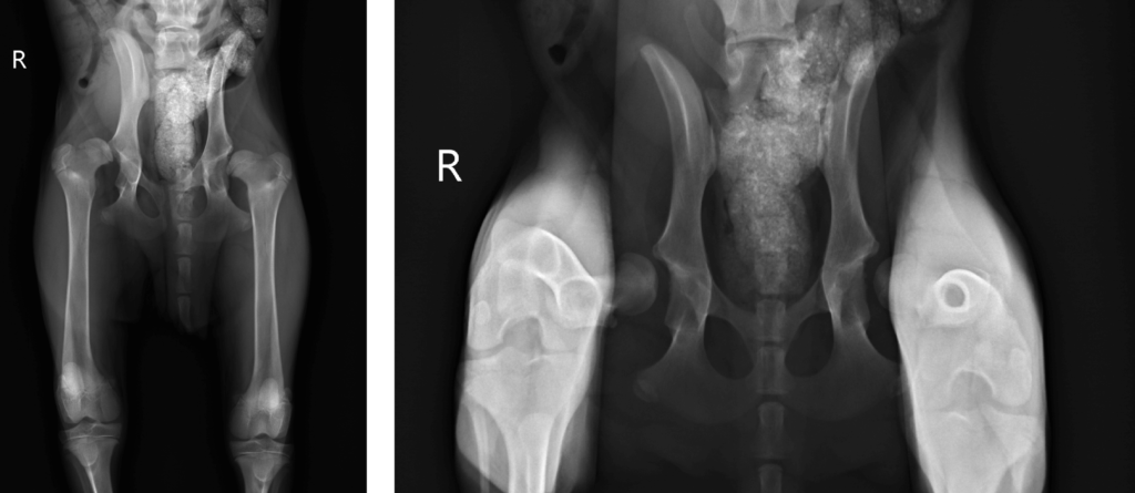
X-ray
On both projections it is clearly visible that the right limb mass is not in the socket. The standing DAR technique should show this: Colour processed blue is a normal hip and red is Frida’s hip when there was no TEP.
This is the reason why the right limb is shorter than the left.

Summary
As seen on the video and x-rays:
Although Frida is doing better overall, the right side, which was not operated on, luxates upwards. A second operation for the defective hip joint is inevitable.
However, if the second operation adjusts the length of the limb and allows the head of the femur to move in the socket, Frida’s quality of life can be restored. Inflammation and pain can heal and healthy movement will be possible.
The second artificial hip joint for Frida will be custom-made for her, just like the one on the left side. Frida’s bones are too thin and the general situation too bad to use a standard prosthesis. The use of the second artificial hip joint will enable a stable position of the right femoral head in the socket. For Frida, after the healing process and exercise therapy, this is the prospect of a life fit for a dog – Full of healthy and painless exercise.
Frida’s story continues!
Visit our social media channels and website to stay up to date.