Functional orthopaedics in dogs – Husky Dante

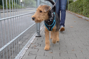
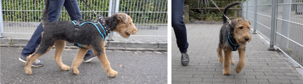
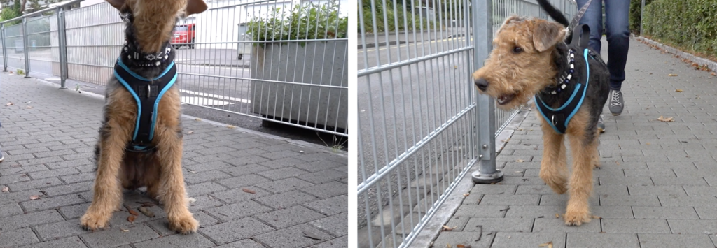
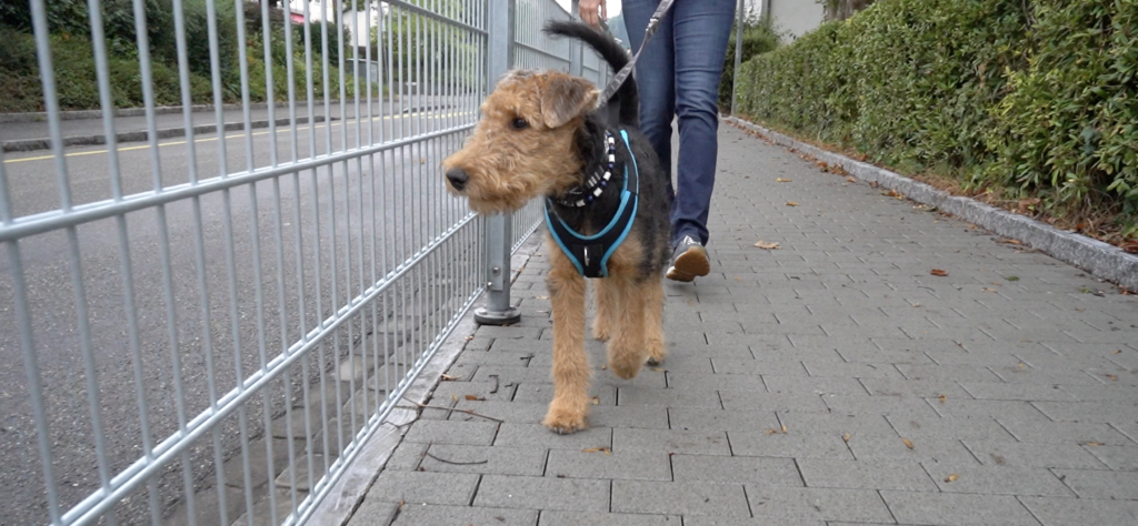
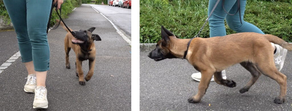
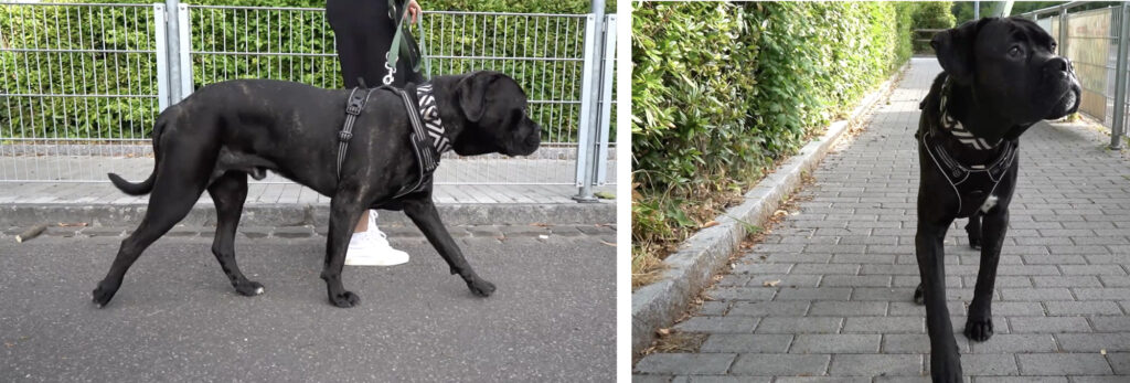
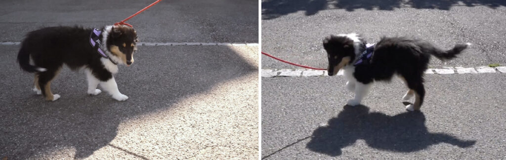
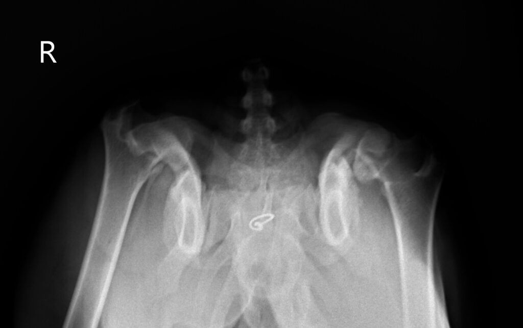
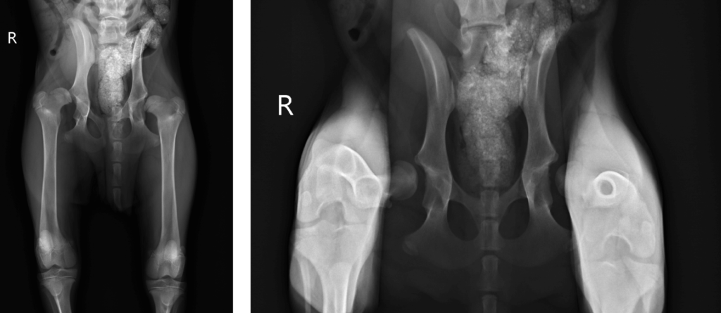
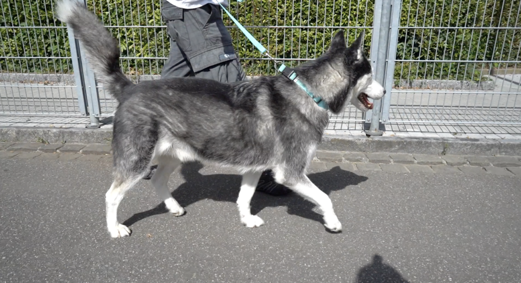
The patient we have the opportunity to present today has been showing right front hang-leg lameness since January. This case not only demonstrates how functional orthopaedics works – it also shows how it can help even “hopeless” patients.

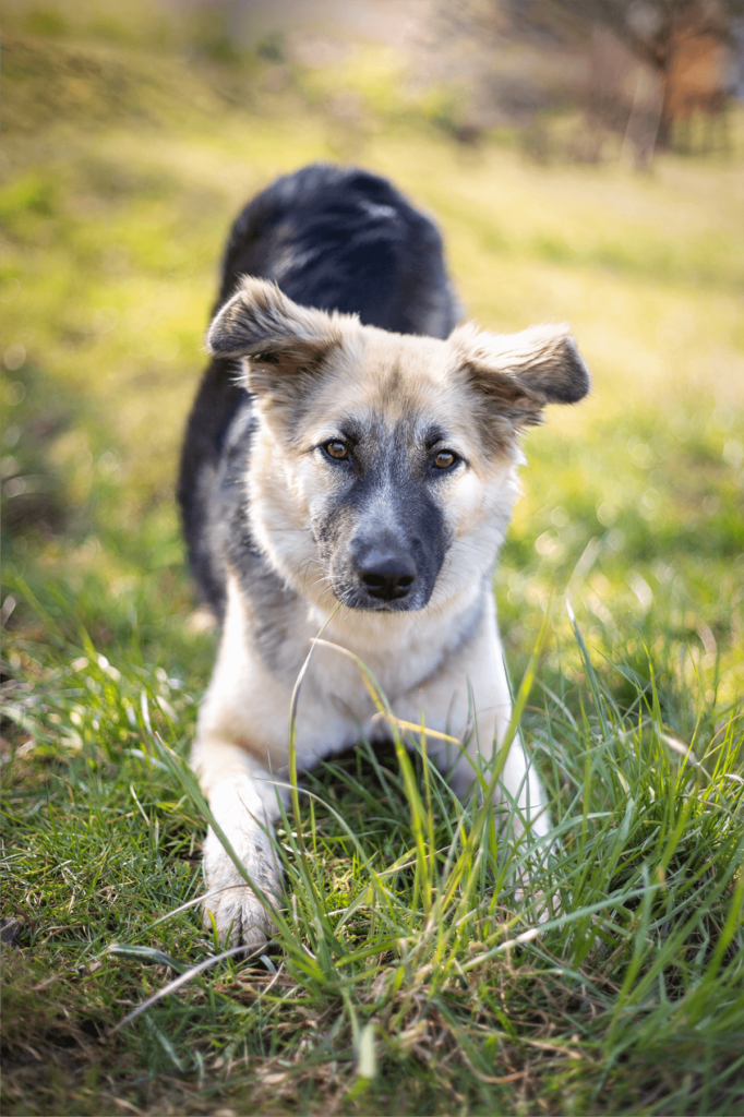
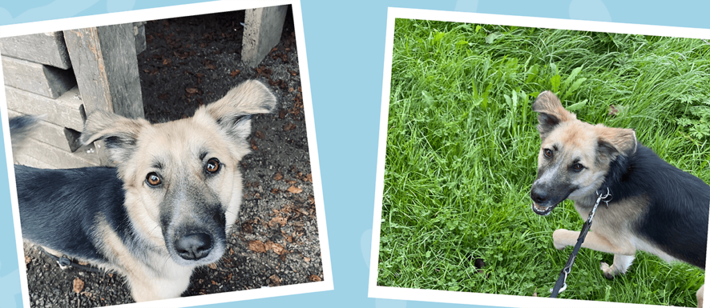
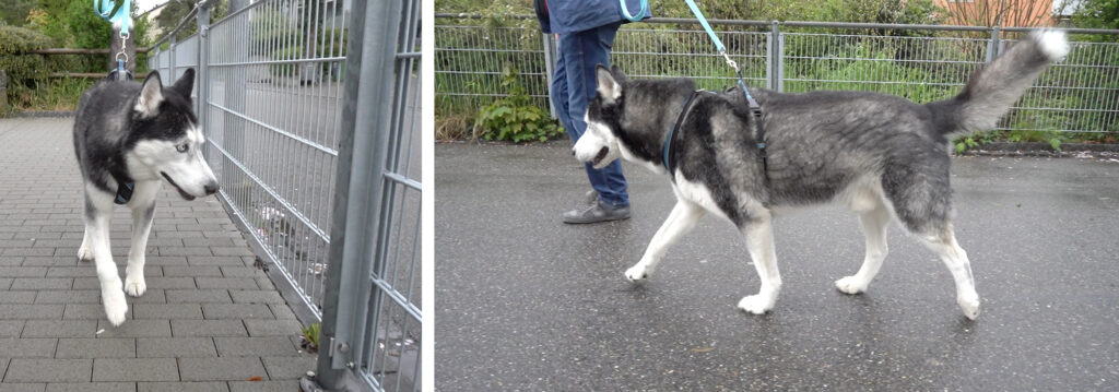
The husky Dante was presented to the in-house vet with a suspected muscle strain. However, anti-inflammatories and physiotherapy prescribed beforehand did not bring any relief. Therefore, he was presented to our practice in April.


Functional orthopaedics

Functional orthopaedics deals with orthopaedic structures and their pathology. In particular, it is concerned with mobility. This includes:

  • The degrees of freedom of joints and limbs.
  • Structural physiological and pathological findings.
  • General (dys)function of the joints as well as
  • Regional orthopaedic structures (such as at the pelvic girdle or spine).
Swingphase
Stancephase
Extension / Flexion
Average Range of Motion

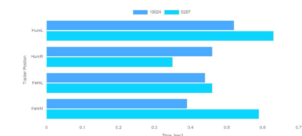
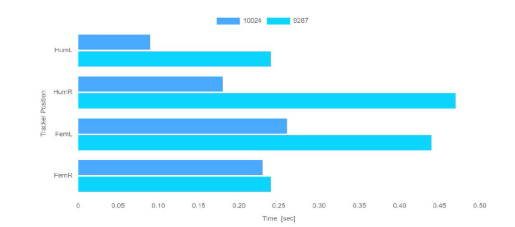
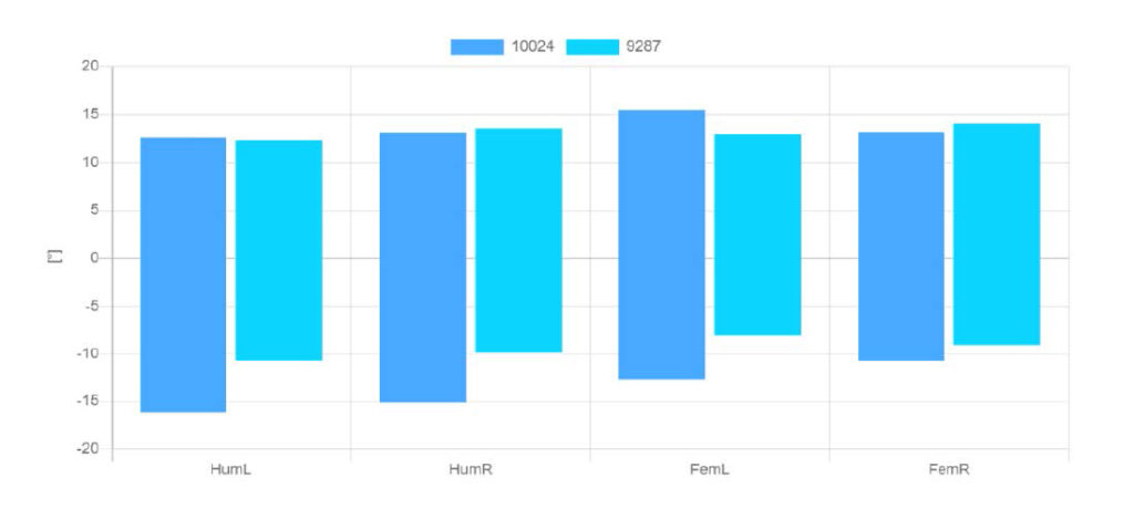
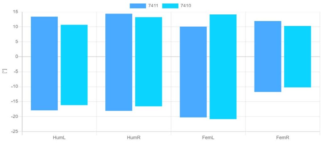
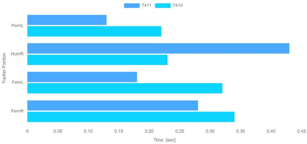
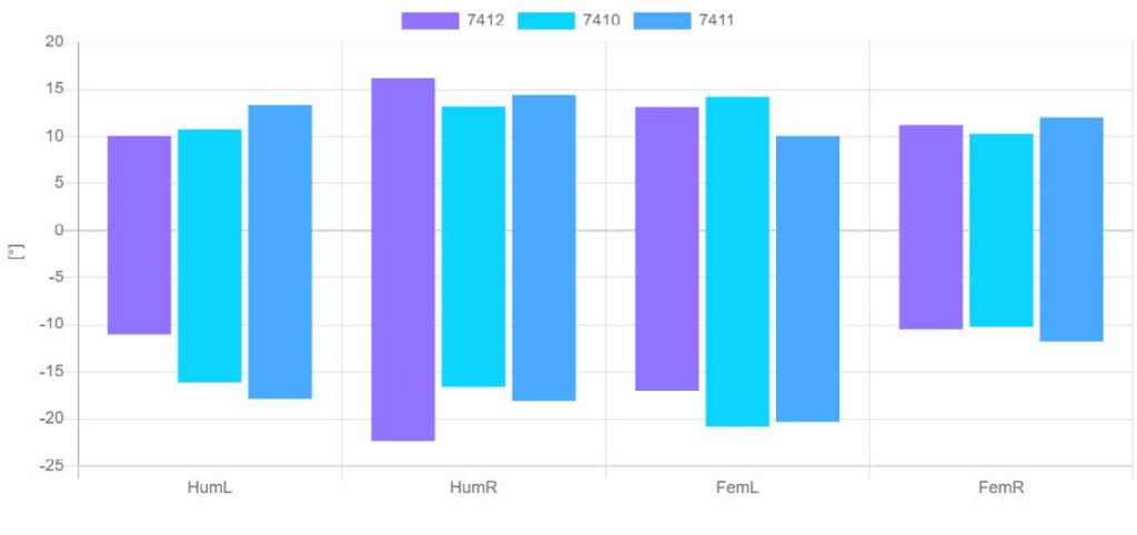
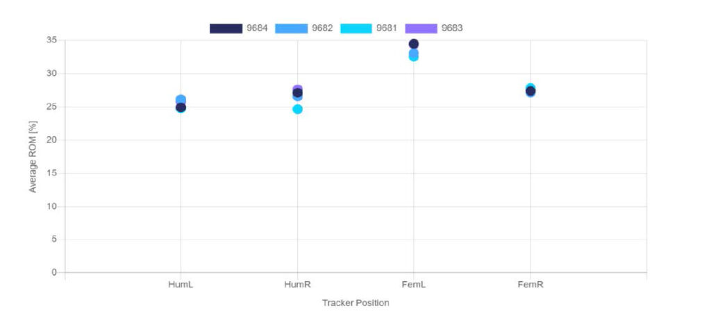
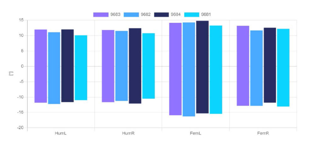
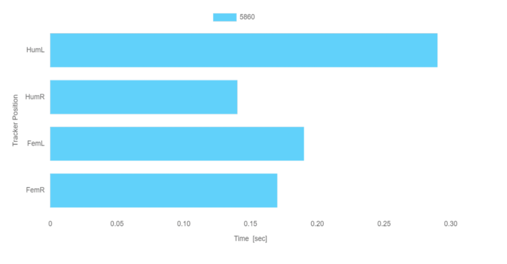
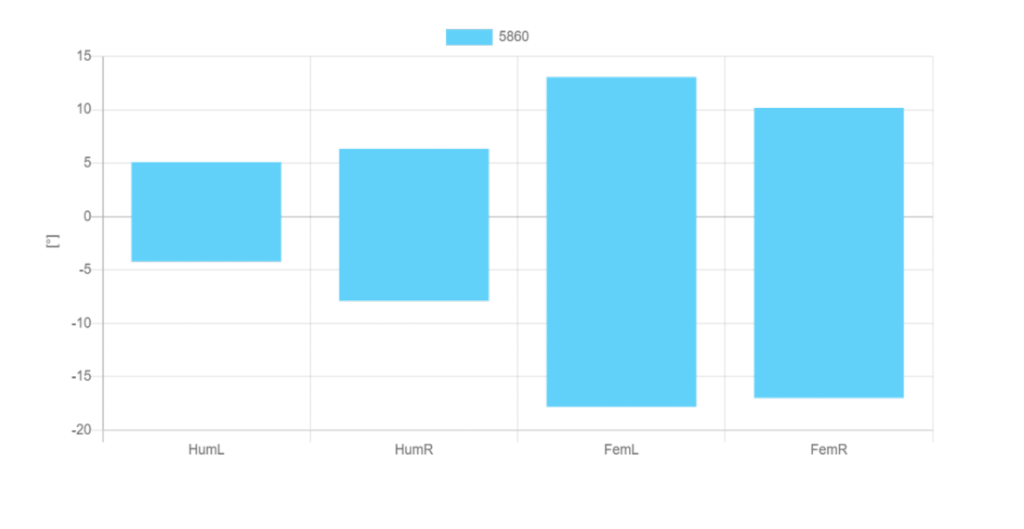
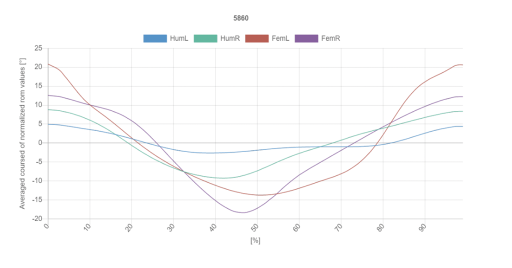
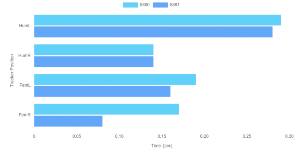
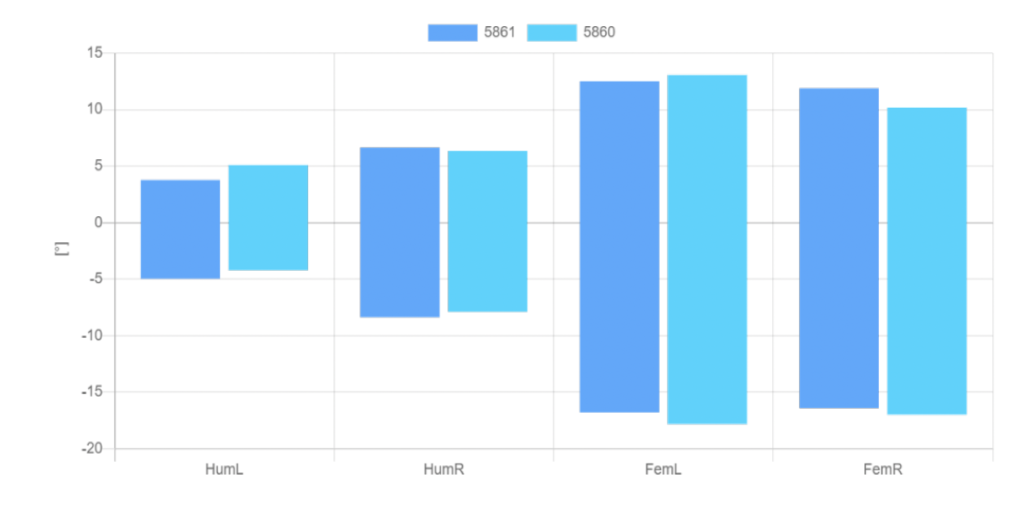
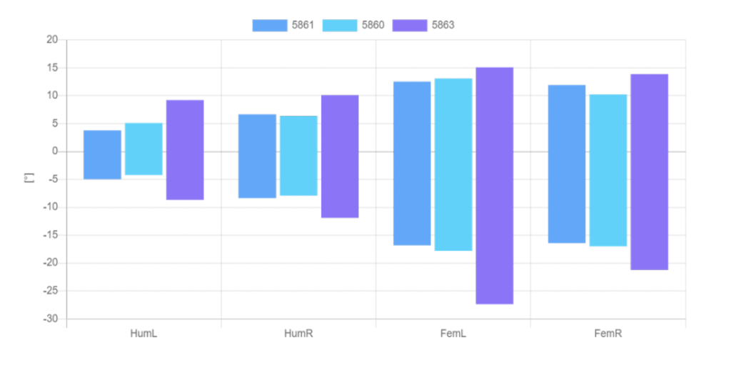
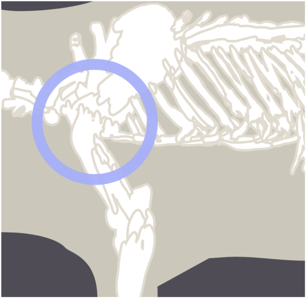
In the case presented here, the dog’s shoulder is the focus. The terms Range of Motion (ROM) and Range of Freedom (ROF) play a special role. They provide information about the function or – in this case – the dysfunction of a joint, whether it is normal, altered, or even physiologically or pathologically conspicuous. 

The examination of functional orthopaedics, like all specialisations, demands a great deal of time and experience. It also requires a lot of time and experience to recognise breed-specific changes in the dogs. The treatment differs greatly, also with regard to the size and mass differences of the patients. In addition to experience and time, a fine instinct is required to meet the individual requirements of the patients.


The goal of functional orthopaedics

In a nutshell: Dogs are orthopaedically examined to detect and treat structural changes.

It is generally assumed that a lameness in a dog must be caused by dysplasia, osteoarthritis or some other defective structure. This assumption is largely correct: 50-60% of lameness in dogs can be attributed to this. The rest of the cases, however, have a functional cause.

It can be stated that in these instances, the statics or dynamics are altered without structural pathologies being present. These cases are usually related to the architecture or anatomy of the dog. 


Where is functional orthopaedics important?

Without going into great detail, this is particularly the case with the

  1. Shoulder girdle and
  2. pelvic girdle.

The shoulder limb mass in dogs is important precisely because, unlike in humans, it is not structured as a rotator cuff, but as a synsarcosis. In concrete terms, this means that the shoulder limb mass is connected to the rib cage by a flange and can therefore slide along it in a relatively unstable manner. ROM and ROF play a special role here. 

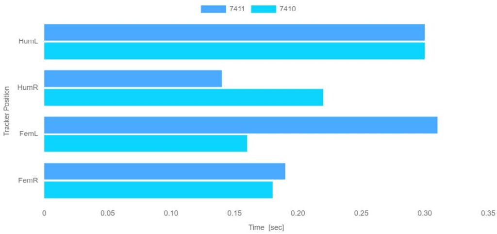
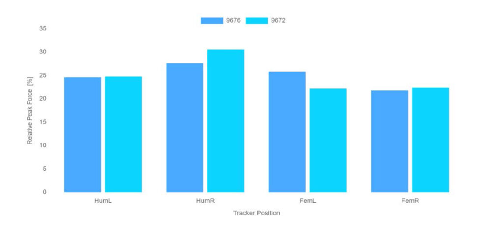
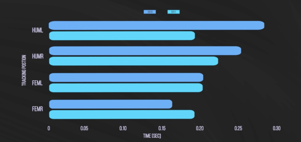
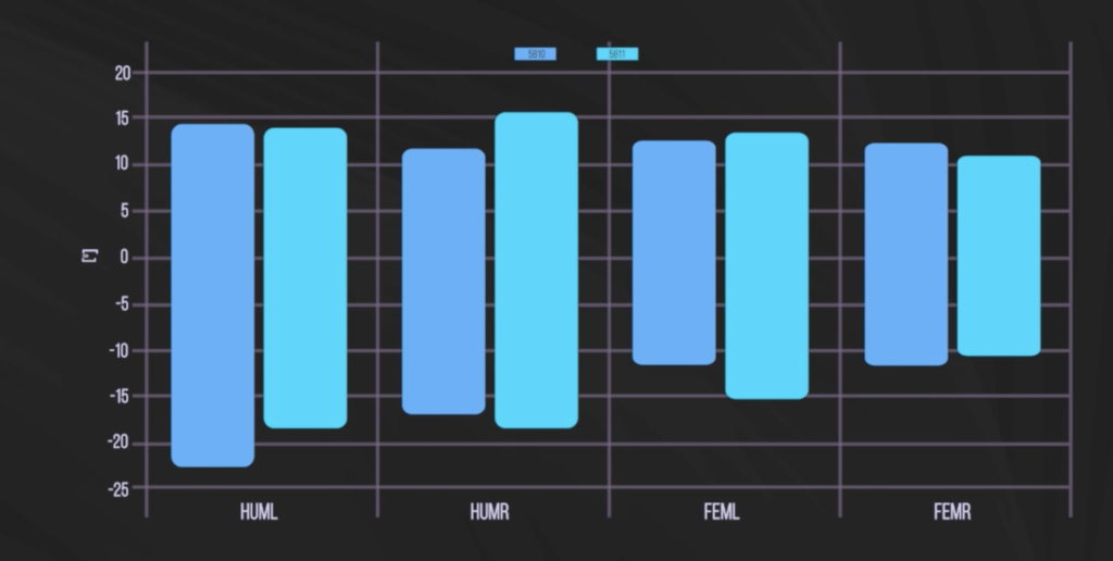
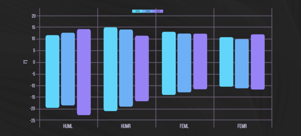
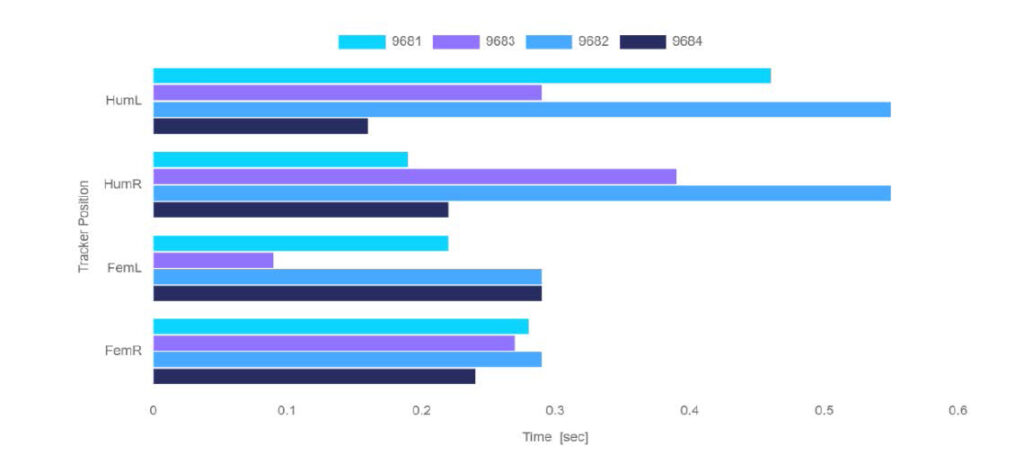
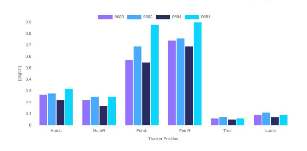
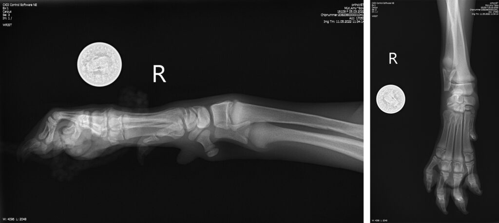
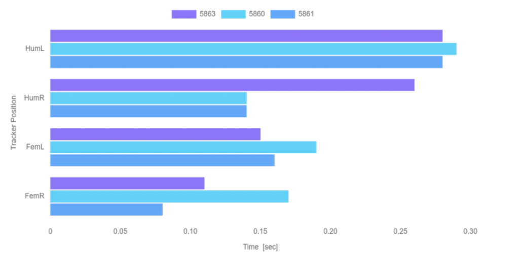
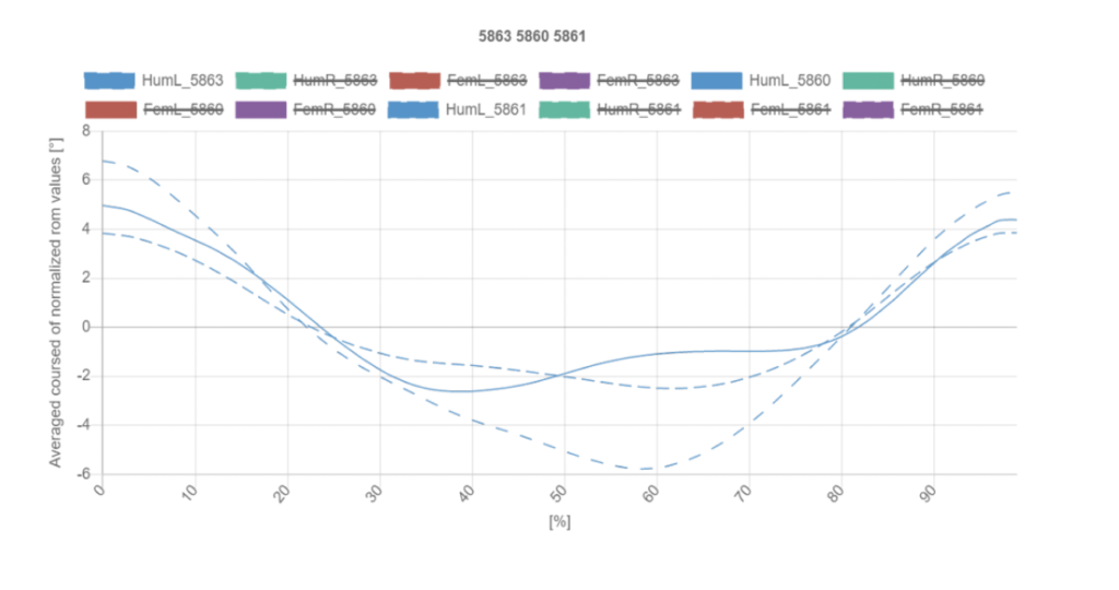
Another point is that many canine shoulder pathologies are cases of soft tissue pathology. As a result, many pathologies cannot be diagnosed by X-ray, ultrasound, MRI or CT. Therefore, in addition to the aforementioned sensitivity of the orthopaedic examination, functional diagnostics are indispensable. This includes gait diagnostics with the “Inertial Sensor Unit” or IMU for short.

The pelvic belt is similar. It is connected to the spine in the same way; the so-called ileo-sacral joint is a 70% connective tissue connection between the iliac-peel joint and the sacrum. Important about the pelvic girdle is the number of exposed nerve endings. Also other receptors that regulate mobility and especially pain nociceptively. Also with this connection, the proportion of soft tissue defines the region. 

In dogs in particular, this area, the cauda equina, is very important for the pathology of the hindquarters. In puppies, the pelvic girdle provides the anatomical structures for hip development; in young dogs, it is significant for training and movement development. And in middle-aged dogs, this is where disc pathologies begin.

It can be stated that this lumbo-sacral instability in connection with the pelvic girdle plays a decisive role in the onset of weaknesses of the hindquarters and pain.


What principles does functional orthopaedics follow?

When we talk about functional orthopaedics, we have to keep the following aspects in focus:

  1. Stimulus-response principle,
  2. Form-function-changes, the
  3. Arndt-Schulze rule and the
  4. Adaptability of orthopaedics.

Let us first look at point a), which is also taught as “actio=reactio”. Specifically in dog medicine: If the dog romps and plays unrestrained and eventually falls violently, this has consequences. These can occur immediately, but also with a delay. It is precisely this that must be taken into account in the case of lameness of the shoulder or pelvic limb masses.

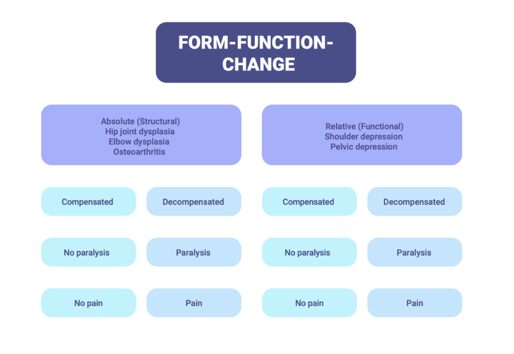
Point b) also comes into play here: a pathology can be structural or functional as well as absolute or relative. In hip joint dysplasia, for example, one speaks of an absolute form-function change. One speaks of relative pathologies in connection with pain in the ISG or a tilted pelvis in the high or low position.

The Arndt-Schulze rule can be explained very briefly. Every physiological movement trains and promotes the stability of the skeletal system as well as the musculature. On the other hand, overstraining movement or overtraining can have pathological consequences. Basically, it is a matter of moderation. This motto is especially important for growing puppies. 

The adaptability of the orthopaedic system can have positive effects. It can mean that the consequences of endurance, strength and speed training are transferred to the orthopaedic system at all times. On the other hand, however, there are also negative consequences.

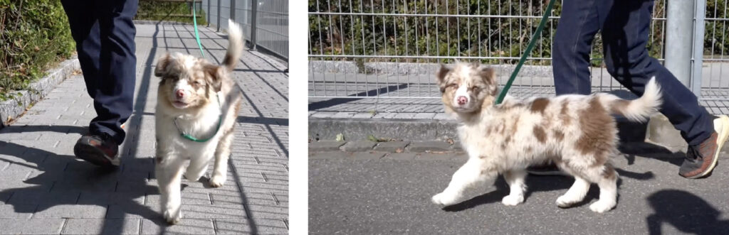
A practical example is the hopping of smaller dogs. Normally this is associated with the dogs’ patella. However, if the habitual patella is not the cause, there is often no explanation. Generally, the bouncing is subsequently treated as a “tic” – which is absolutely not true. After all, the dog does not show any orthopaedic compulsions or comparable behaviour. Functional orthopaedics provides information here and attributes the problem to the biomechanics of the pelvis. Through romping, playing and uncoordinated movements, these biomechanics changed, while the growth plates are still open due to the dogs’ young age. These changes are subsequently reflected in the pelvic limb mass. Variations in statics result in variations in dynamics. Bouncing is a countermeasure designed to compensate for specific differences in frequency, step or stridelength. A suitable analogy is a differential in automobiles: This compensates for differences in wheel revolutions of the axle, for example in curves.

It is also worth mentioning at this point, in addition to functional orthopaedics, the role of functional neurology – also in relation to the bouncing of smaller dogs. More of this at another time.

In summary, it must be stated that functional orthopaedics and thus statics and dynamics must increasingly find their way into veterinary orthopaedics – with the inclusion of biomechanics and its rules. Functional diagnostics will provide the necessary basis for the treatment of non-structural disorders.


You can also watch a video of Dante’s case:


For more information on kinematic gait analysis and the correct build-up of puppies, take a look at our book “LUPOMOVE® PUPPY – THE INNOVATIVE DEVELOPMENT AND PROPHYLAXIS PROGRAMME FOR PUPPIES”.

Case study of Daisy, 18 week old Airedale Terrier

Initial situation

Airedale Terrier, 18 weeks old, missing toe.

The dog was presented to the practice for a prophylactic examination and for the right rear limb which was missing a toe. This fell off, probably due to necrosis. On movement, we see that the dog has a conspicuous gait on the right rear.

More precisely, this means that the axis from the hock joint downwards turns inwards and causes a slightly circular gait. The activity of the hindquarters seems to be reduced, it also goes into a left outward stance, at times also into a right outward stance. 

Viewed from the front it is noticeable that the mobility of the left shoulder phalanx is altered compared to the right. 

Viewed from the side, the forehand appears, at first glance, to be relatively ground covering. The hindquarters sway a little due to the position to the right and left outside, which makes the mobility and activity there seem reduced. 

All in all, the changes are only slight; only the limb position at the rear right from the hock down towards the toe is noticeable.

Diagnosis

The diagnosis revealed an altered croup musculature on the right side, furthermore the mobility is different on both sides of the pelvis. Especially the gluteal muscle has a different shape on the right side. The positions of the hind limb are also different. Seen from above, the S-shaped curve of the spine stands out; in front of the pelvis there is a slight left torsion. In the middle, the spine twists and continues to the right.

This is followed by the unevenly positioned shoulders: more turned out on the right side, while the left side appears flatter and more choppy in movement. These are the most important findings.

Apart from this, the position on the left rear side is also altered: The puppy is taking weight there, especially on the third toe, and turns it in to compensate on the right side. We see that the shoulder on the left goes forward in one position: an axial cranial translation. Meanwhile, in the pelvis there is an abaxial translation, i.e. a low position, which is also slightly twisted backwards.

It is likely that the toe fell off due to pressure necrosis, caused by another puppy or the mother. The blood circulation was disturbed by lying on the hindquarters for a long time.

First Functional Movement Measurement with LupoGait®

The first functional movement measurement with LupoGait® reflects the diagnosis. Light blue here represents the initial measurement, dark blue after treatment and rotation of the pelvis.

Especially the stance phase at the back left shows a clear improvement after the rotation of the pelvis. But also the flexion and extension of the forehand could be clearly improved.

This means that the puppy has performed a so-called pullmove, i.e. a pulled movement. This explains the less active hindquarters. Through the treatment, the hindquarters as well as the forehand could be relieved accordingly; the hindquarters could be led into a normal traction.

Stance phase
Extension / Flexion
Swing phase

Renewed treatment

As the result was not yet satisfactory, the left shoulder was then manipulated or orthopaedically treated manually so that it was parallel to the right shoulder.

Control measurement with LupoGait®

This led to an increase in the stance phase of both the right and left shoulder, as the control measurement with LupoGait® showed. The stance phase of the hindquarters could be practically normalised. The extension and flexion show an adjustment in this respect: Although the pelvis is manipulated and the shoulder symmetrical, the puppy is still in a compensatory posture on the front left as well as on the back right. This can be remedied by constructive therapy.

The gyroscope readings – in maximum and variance – are now symmetrical, confirming the suspicion of the pull move of the forehand compensating for the hindquarters.

Stance phase, second measurement
Extension / Flexion, second measurement
Variance of Differentiated Gyroscope Signal

Concluding remarks

Following this consultation, it was noted that the dog had an excessive EIK score. “EIK” refers to the energy intensity coefficient; its increase explains the puppy’s increased growth rate. 

The EIK for Airedale Terriers is 650 to 700 grams. At 850 grams in our patient, we ordered a change in diet to avoid growth problems. Already the puppy looks very big, especially compared to his littermates. The dog is fed with barf – we have reduced the meat ration from 80 to 60% and increased the amount of vegetables. We hope that this will regulate the EIK, which we will be able to check in a control examination in three weeks. 

Watch the case of Daisy also as a video:


For more information on kinematic gait analysis and the correct build-up of puppies, take a look at our book “LUPOMOVE® PUPPY – THE INNOVATIVE DEVELOPMENT AND PROPHYLAXIS PROGRAMME FOR PUPPIES”.

Case study of working dog Pico, 14-week-old Malinois

Initial Situation

The puppy Pico was introduced to our practice so that the build-up to a working dog could proceed without problems. Another goal was to develop the training more efficiently.

First examination

The first examination already showed a complex gait pattern. Pico cannot walk straight, the right hind quarters always go into a right outward position. In addition, the right hind limb bends. In movement there is a clear, strong swaying to both sides. Additionally relevant is a tucked up croup, a tendency to limp front-left as well as the gait of the left shoulder limb in general, which is broad-legged to circular. 

Basically, the gait is irregular and abrupt, the hindquarters are heavy and not symmetrical.

This gait pattern is special, but unfortunately not rare in the puppies that come to the practice. It is always pathological and forms the basis for a pass gait. This pattern forms the centralisation of a later gait pattern if it is not recognised and corrected early. 

Orthopaedic examination

The orthopaedic examination shows symmetrical knees and ankle joints. The left femur is displaced downwards and backwards, which clearly restricts its mobility. In addition, there is an abaxial rotation of the left pelvis, which corresponds to a pelvic low position there. The sacral region also has reduced mobility. The sensitivity of the examined anatomical regions is remarkable. Last but not least, the left shoulder is also limited in its mobility, and its axial rotation also stands out, which causes the limping there.

The findings of the orthopaedic examination were supported by the kinematic measurement.


Kinematic measurement


The kinematic measurement shows a difference in stride length between the left and right pelvic limb of 10 cm, which is striking. Furthermore, the stance phase and the range of motion show only slight differences, while the PVF in the hindquarters shows no difference at all. The left shoulder has a reduced acceleration, which indicates a pull move to compensate for the instability of the hindquarters. In general, altered values are present but not dramatic. There is no evidence of structural pathology or hip dysplasia. Therefore, an X-ray study was not performed.

Stance phase, initial measurement
Maximum amplitude of the gyroscope signal, initial measurement
Vertical peak force, initial measurement
Step length

Orthopaedic-manual treatment

Orthopaedically, the malposition in the pelvis now had to be corrected manually.

Kinematic control measurement

After the manual treatment, Pico was again kinematically measured and controlled.

After the treatment, a symmetrisation of the pelvis kinematically took place; however, this was not completed because the malposition in the pelvis on the left side had already led to compensation of the movement in the left shoulder.

Overall, there are clear improvements, so that the further build-up could be planned.

Stance phase, second measurement
Maximum amplitude of the gyroscope signal, second measurement
Peak Vertical Force, second measurement

Visual results

The visual gait pattern after the orthopaedic-manual treatment is a clear improvement: Pico shows good movement in the trail, the instability of the hindquarters has practically normalised. The swaying as well as the buckling of the hindquarters is no longer visible and the lameness of the left shoulder disappeared. The whole movement now appears round, harmonious and fluid.

Folgetherapie

Now the targeted development of the movement and the musculature can begin. Without this correction, Pico would only have built up the musculature incorrectly and, above all, too little and the altered gait pattern would have manifested itself peripherally as well as centrally in the motor cortex in the brain. This would have been detrimental to Pico’s use as a working dog.

Watch Pico’s case also as a video:


For more information on kinematic gait analysis and the correct build-up of puppies, take a look at our book “LUPOMOVE® PUPPY – THE INNOVATIVE DEVELOPMENT AND PROPHYLAXIS PROGRAMME FOR PUPPIES”.

Case Study of Camila, 10 week old Australian Shepherd Puppy

Initial situation

Camila, Australian Sheperd, 10 weeks old, has a left front limp.

Camila fell several times from an elevation; the first limp normalised after 2 hours, but after the second fall the irregular gait persisted.

Therefore the owner came to see us the following day.

Examination

The puppy walks relatively tight in the forehand. Especially the left shoulder joint is turned into an inside position, which causes a toe tight gait. Also the limb axis is oblique. At the top it is slightly turned outwards, at the bottom it is turned inwards. 

The space grip of the left shoulder limb is reduced, as also visible on the movement guided from the side. The movement of the left shoulder limb appears broken, the hovering phase is shortened.

Camila walks with the hindquarters out to the left, which reinforces the lameness and limping on the left front. 

As no structural changes to the bones were apparent, a radiological study of the left shoulder limb mass was not performed.

Kinematic study

We used the measuring jacket and our gait analysis system to check the treatment and quantified values. You can see the findings in the following graphs.

Light blue are the findings before the orthopaedic biomechanical kinematic controlled manual treatment, dark blue those afterwards.

It is noticeable that especially the stance phase as well as extension and flexion have improved significantly. Also the gyroscope values; the angular accelerations of the limb have changed. After the orthopaedic treatment, the findings are symmetrical. In concrete terms, this means that Camila can put relatively normal weight on the left shoulder limb again.

Stance Phase
Extension / Flexion

The orthopaedic biomechanical manual treatment regulates the statics and dynamics of the limbs and the spine. The goal is the physiological (normal) function of the joints as well as goal-directed movement. 

Furthermore, this movement should be possible without inflammation or pain. A limp in a dog is always an indication that the movement is not normal; due to faulty mechanics or inflammation and pain.

In Camila’s case, we set up a movement plan for 14 days following the treatment and then checked her values again (purple).

The video sequence shows a smooth, round movement. Both the shoulder and pelvic limbs moved actively and expansively, the position of the left shoulder limb mass was practically normal. The only noticeable thing was that in the sitting position, the left shoulder limb mass was placed slightly forward. 

Our new kinematic measurement (violet) showed an altered extension and flexion. It has to be taken into account that the puppy grows daily, so a slight discrepancy in the values of individual features is to be expected.

Extension / Flexion, third measurement

In conclusion

Unfortunately, it happens very often that a puppy falls off sofas, boxes or small walls. The coordination is still lacking in the young dog, the stability and strength of the pelvic limb and the musculature is not yet developed.

If these cases are not orthopaedically biomechanically examined and treated, this can have major consequences, especially in the hip area.

But shoulder pathologies and lameness are also common; they can even often persist despite treatment. The reason is the altered biomechanics of the shoulder – the range of freedom. This must also be put in the context of OCD – Osteochondrosis.

We will dedicate a separate webinar to this topic: Prevention with the LupoMove® programme focusing on shoulder and hip disorders in puppies and young dogs. For more information on cartilage regeneration, recommend our book “LupoMove® sports, works and family” for a detailed explanation.

Watch Camila’s case also as a video:


For more information on kinematic gait analysis and the correct build-up of puppies, take a look at our book “LUPOMOVE® PUPPY – THE INNOVATIVE DEVELOPMENT AND PROPHYLAXIS PROGRAMME FOR PUPPIES”.

Cane Corso, 9 months old – Capper Case Study

In this case we present a Cane Corso, 9 months old. He walks stiffly in the hindquarters, shows a painful pelvis and trembling forelegs. He was presented for a second opinion at the practice of Dr Blättler-Monniers.

Initial situation

Capper walks stiffly in the hindquarters and shows pain in the pelvic region. In addition, he trembles in his front legs.

The owner has already been to the PTA who advised to wait with the treatment for the time being. In addition, a shell compound was given. When there was no improvement in the gait, the dog was brought to our practice for a second opinion.

Gait pattern

Capper’s gait pattern is very stiff. He shows reduced bending in both knees. In addition, there is toe widening in the left pelvic limb. On the right side, the pelvis is low, which causes an obliqueness. The left shoulder is less spacey. On the move, Capper is in a right outset position and traverses over the right shoulder.

From the side, a slope leg lameness can be seen in the right and left pelvic limb. In addition, both pelvic limb masses are in an ankurvature and are placed below the centre of gravity. The bending of both knees is reduced. The back shows kyphosis. 

In general, the hindquarters show little drive; Capper performs a pull move and pulls mainly with the forehand. The left pelvic limb is passively pulled and appears even more ponderous.

Gait pattern on the volte (circle)

On the volte, Capper looks similar: His movement is ponderous. The drawn up back has an even greater effect under these circumstances. In addition, the slope leg lameness has increased on the right as well as on the left. We are talking about turning pains here.

Especially the left pelvic limb mass seems to be strongly reduced in movement, the toe widening has also increased.

Last but not least, the pelvis tilts when sitting. The left side appears much more unstable.

Diagnosis  

Capper shows bilateral hip dysplasia: moderate on the right side, severe on the left.


Kinematics


The kinematic data of the LupoGait® measurement supports the diagnosis: the reproducible movement features also indicate hip joint dysplasia.

Stance phase
Variance of the differentiated gyroscope signal
Range of Motion
Extension / Flexion

Therapy (short-term)

In the short-term therapy, the pelvic axis was treated orthopaedically-manually. Furthermore, bednivetmabum was prescribed as injection therapy until the pelvis had grown out.

Depending on the course of the clinical symptoms, an individually tailored hip joint prosthesis (custom-made TEP) is indicated.

Watch Capper’s case also as a video:


For more information on kinematic gait analysis and the correct build-up of puppies, take a look at our book “LUPOMOVE® PUPPY – THE INNOVATIVE DEVELOPMENT AND PROPHYLAXIS PROGRAMME FOR PUPPIES”.

Hidden factors in the treatment of supporting leg irregularity: Case Study Sammy

Puppy, 9+ weeks old, supporting leg irregularity of the front-right limb since one week.

This real-life case is presented by the World Veterinary Organisation for Kinematically Controlled Orthopaedics, Neurology and Rehabilitation. It´s founder and chairman of the board is Dr. med. vet. Patrick Blättler Monnier. 

Initial problem 

The little puppy moved in with the family only a week ago. Shortly before that, she had come from a breeder in the north. She was intended as a bitch for breeding.

The day after, Sammy started to limp. The owner made an appointment for an orthopaedic puppy consultation with us.

The supporting leg irregularity in the front on the right is clearly visible. The toe axis there is turned outwards, there is toe widening. There is also swelling of the forefoot joint. The axis of the right shoulder phalanx seems to be turned obliquely inwards, moreover, two toes are almost dropped on one line. The narrow position of the shoulder limbs is striking, as is the drawn up back and the very steep position of the hindquarters.

Orthopaedic functional examination

In addition to the angulated toe axis and the swelling in the forefoot joint, it is particularly noticeable that the extension of the right shoulder is clearly reduced. The puppy reacted with defensiveness to the examination. The left shoulder could be moved normally. A verdigris fracture cannot be excluded. 

Therefore, an X-ray of the paw is taken and a kinematic examination is carried out as part of the LupoMove® Puppy programme.


X-Ray diagnostics

Both x-rays of the paw showed no abnormalities. A fracture could be ruled out and no other structural changes were found.


Kinematics 

It is remarkable that the stance phase of the front-left leg is 0.29 sec and of the front-right 0.14 sec. The difference is 100%. 

The difference in extension/flexion between right and left was also considerable, caused by the unloading posture of the right limb and the overloading of the left. 

The flat “average step” curve of the left limb compared to the values of the other limbs, also a result of overloading, is also striking.

After the diagnosis, the tarsal joint and the rotation axis were treated orthopaedically-manually and then measured kinematically. The direct difference can be seen in the light blue results before treatment compared to the dark blue results afterwards.

The kinematic mobility of the shoulder was partially restored, but not to the desired extent.

Stance phase, second measurement
Extension/flexion, second measurement

Therefore, the findings from the video analysis came into play and were included in the treatment.

In the second orthopaedic-manual treatment, the left shoulder limb was treated manually, as well as the hindquarters, i.e. the pelvis and both hind legs. Again, a kinematic control was carried out.

This time, the result was satisfactory.

The values of the third measurement are highlighted in purple. It shows that the stance phase at the front right has clearly increased. It increased from 0.14 sec to 0.26, on the left side it is only slightly higher at 0.28. The puppy’s gait is almost symmetrical again. The extension/flexion also normalised, the average ROM evened out. This can be seen well in the development of the “average step” curve of the left shoulder limb. 

Stance phase, third measurement
Extension/flexion, third measurement
Average step-curve, third measurement

Conclusion

The acute appearing limp of the right shoulder limb was directly related to the left shoulder limb as well as the hindquarters. Both changes are semi-acute and could not have been distinguished without the kinematic examination. 

The limp, which could have been interpreted as a consequence of the malloading and overloading of the right shoulder limb, is only part of the problem. Not only the right shoulder limb, but also an orthopaedic malposition in the pelvis and the left shoulder limb affected the puppy’s gait.

Without the kinematic treatment and LupoGait® the examination would not have been so efficient; this is precisely the advantage of the orthopaedic-kinematically controlled puppy consultation.

What could have happened – The imprinting and conditioning of altered movement and pain processes in the puppy.

This patient came to us because of a supporting leg irregularity on the right front. Unfortunately, limping is not uncommon at the age of 10 weeks – but without the kinematic, differentiated diagnosis, the puppy’s altered gait pattern, especially the incorrect and overloaded posture, would have remained undetected.

The pain process and the altered movement can quickly be conditioned.

We would like to explain what the consequences would have been without the treatment.

Young dog diseases such as hip and elbow dysplasia, osteochondrosis or panostitis are epigenetic diseases. This means they result from genes that are encoded and expressed by environmental factors. This is where the altered movement of the puppy arose.

The triggering environmental factors that mobilise the genes are not uncommon.

All the more reason to present changes in gait directly to a professional, orthopaedic puppy consultation. 

Here we determine these external, triggering environmental factors in order to reduce and minimise them. In this way we strengthen or restore the health of the puppies.

However, there are other points that need to be taken into account in such gait changes. These are above all

  • Conditioning of the altered gait with consequences for the orthopaedics
  • Imprinting of the pain process and thus the behaviour.

The second point in particular is very important, because it must be assumed that the psycho-social part of the imprinting in the puppy can be strongly and potentially negatively influenced. Pain is a stressor in the puppy; in our opinion one of the most powerful, often severely affecting behaviour, but also general health.

From a behavioural perspective, pain can make it difficult to interact positively with other dogs and puppies, as well as with the owner or strangers. The main behavioural modalities are fear and aggression, but restraint to apathy and lassitude can also result. A puppy loses his open and fun way of exploring and investigating. He is less carefree. He can withdraw, seem reserved.

The stressor “pain” also has an influence on general health, resistance and the immune system. Stressed dogs, and those in pain, fall ill significantly faster. Parasitoses occur more often. 

A good example is Giardia: these bacteria are only facultatively pathogenic. Healthy puppies do not get sick from them. Stressed dogs often have prolonged diarrhoea and are more often resistant to therapy.

The last point we would like to make is about cognitive learning. A puppy in pain is generally slower to learn and less likely to pick up new activities. 

The puppy may also be generally withdrawn, or the dog may react with behaviour similar to stimulus overload. These psycho-social behaviours should always be investigated for pain as a cause. These behaviours strongly influence the imprinting, conditioning and learning curve of the puppy and young dog.


Unwanted imprinting of movement

The altered gait pattern also has a strong influence on general movement, balancing and therefore also on statics and dynamics, in other words: the biology of orthopaedics.

Movement is first controlled and regulated in the periphery at the bone, joint and musculature. This is where we find the muscle and joint receptors. The information generated there is passed on centrally via the spinal cord and brain stem. This data is processed in the midbrain and cerebellum before it is transported to the cerebrum. Here, the movement is consciously coordinated and responded to. This information is then passed back to the periphery via the data highway and results in action. 

If we show such disturbances in the puppy in the 10th week of life without treating them purposefully, we lay the foundation for gait and movement abnormalities as well as pain. Neither vets nor dog owners are sufficiently aware of this fact. Only with movement diagnostics, in particular with LupoGait®, can these pathologies be detected.In order to counteract these pathologies, a professional, ortho-kinematic puppy consultation, such as within the framework of LupoMove® Puppy, is the only variant to treat disorders at an early stage and to prevent the consequential damages shown.

Watch Sammy’s case as a video:

Frida’s Story – A new hip for Frida

A street dog fights hip dysplasia and pain

Frida’s journey begins as a street dog in Romania. As a young puppy, she was found in a rubbish bag. An international animal welfare organisation rescued her and brought her to Germany to her new family. Her new-found happiness lasted only a short time, however, because soon after her arrival she developed difficulties walking. Shortly after, she was no longer able to stand.

Her condition was diagnosed as bilateral hip dysplasia;

her condyle didn’t sit properly in the socket, often popping out. The cartilages rubbed against each other, which after a while destroys their natural shock-absorbing function and can lead to hip arthritis. All this caused Frida severe pain. A juvenile pubic symphysiodesis, a minimally invasive procedure on the growth plate, was to serve as a temporary solution. Frida was still too young to have an artificial hip joint.

Diagnosis and symphysiodesis

Situation
Frida’s case shows well the typical problems of small and growing dogs. A week ago, Frida fell out of the car; since then she has been lame in the right rear and has difficulty standing up. Because of this, and for general prophylaxis as part of the LupoMove Puppy programme, Frida was presented to the practice of Dr. Blättler-Monnier.
Initial visual assessment 
The clinical picture is already visually multifaceted: Frida’s back is tucked up, especially towards the middle, and her hindquarters are very unstable. The pelvis swivels in both directions when walking, both hindlegs are swerving backwards in a circular motion. In addition, the hock joint is noticeable, which is directed strongly from the inside to the outside.
When Frida sits down, she positions the forelimbs in a line, the pelvis is tilted and both hind legs are turned out to the left.
All in all, Frida appears unstable and uncoordinated. The front half of her body is almost inconspicuous – but the back looks quite different. It looks like she is in a lot of pain.
Orthopaedic examination
The orthopaedic examination revealed a conspicuous decrease in the pelvic muscles of the right side.  The deficit was 2cm: We can speak here of a strong atrophy, an underdevelopment of the musculature.
The mobility tests carried out, the Ortolani test and the Barthens test, were both positive. This also indicates instability of both hip joints, which is also associated with pain.
Both knees behaved normally; the test to detect torn cruciate ligaments was negative. This would have been unlikely, but possible.
We also performed a drawer test, a TPA compression test and a TPA parvo test with external rotation. The ankle joints were also unremarkable. In the lumbar spine, the strong, long extension of the back due to underdevelopment was particularly noticeable. The lumbar spine is limited in its mobility.
Neurological examinations 
The neurological examinations did not reveal any abnormalities.
 After visual and orthopaedic-neurological examinations, we can make a tentative diagnosis of hip joint dysplasia. This will now be examined in imaging procedures.
X-ray
For this purpose, Frida is sedated in order to take X-rays in a stretched position. These are imagined below.

It is easy to see that the hip does not move in the socket in an extended position. Also in a frog position the hip is loose on both sides.

The third picture shows a distraction technique. The hip is spread and the looseness is tested at the same time. In Frida’s case the looseness is 1, which indicates a very loose hip.

Diagnosis and outlook

As a result of the diagnosis, we discussed the clinical picture and the resulting options with the owner. The owner immediately decided against euthanasia. 

She was clearly informed that the further treatment would have to be divided into two steps. On the one hand, a short-term therapy, the symphysiodesis, to support Frida’s growth in the next months, to reduce her pain and to allow her a more normal socialisation.

In the medium term, two artificial hip joints will have to be inserted afterwards.

Furthermore, the owner was shown the exact costs and therapeutic consequences for the dog, as well as the duration and type of rehabilitation and regeneration. 


Symphysiodesis – Frida’s first operation

Following the examinations, Frida immediately underwent a juvenile pubic symphysiodesis. This means that the pubic symphysis is surgically closed at an early stage. The technique itself is a relatively new procedure that was first successfully applied to guinea pigs and has also been successfully applied to canine patients for some years.

It is a prophylactic preventative surgery that is used primarily in growing dogs that show a great tendency to hip dysplasia and loose hips.

Laser surgery is used to cause a portion of the pubic symphysis to ossify. As a result, the lower, rear part no longer grows, while the upper part continues to form. This results in a better roofing of the femur bones than would have been the case without therapeutic measures – the stability of the joints is increased.

The symphysiodesis was successful, as was the post-operative check: Frida no longer walks with a severe limp.

Outlook
However, in Frida’s case, this preventive treatment is not enough to completely eliminate her HD. Despite the success of the surgery, it only provides a means to allow Frida to grow normally without pain. She should be able to move normally, at least with only a little lameness, for the next few months until she is fully grown. However, without major surgery, Frida’s hip will never be able to function like a normal one.
At a later date, when all the growth plates are closed, Frida’s treatment will continue.

Frida’s custom-made TEP – the left hip joint

At a later stage, when all the growth plates are closed, Frida’s treatment will continue.

The initial situation

Frida is now in Dr. Blättler-Monnier’s practice. It is early autumn 2022, 7 months after her successful symphysiodesis. She is again showing pain and lameness, especially on the back left side, after lying down and moving around, but also after playing with other dogs.


New hip joint from 3D printing

The reactive hip is the cause of Frida’s problems. Frida is now older – so an artificial hip joint for the left side was considered. The orthopaedic examination only confirmed this. However, no simple, cementless TEP (total endoprosthesis) was to be inserted. A 3D-printed TEP was created based on the bone and pelvic structure. 

For this purpose, Frida was first referred for a CT study. The data collected was processed and used to create a model corresponding to the hip prosthesis. The model was tested to see if it could be attached to the bone and work correctly from a biomechanical point of view. The tests were successful – a prosthesis for the left side was created, which was inserted before the end of autumn 2022. Physiotherapeutic measures following the operation should help her to walk evenly.


Frida’s fight against hip problems continues: second operation necessary

Frida came through the operation well overall, and the control of the newly implanted hip was also successful. In January of this year, Frida again developed difficulties when walking: she again showed an irregular gait; in addition, she has pain in her right hip.
The left leg turns outwards and Frida is also sensitive at the back left, at the operated hip. The owner came for an orthopaedic re-evaluation. 

In movement, the left, operated side turns outwards, while the right side shows a clear slope leg lameness. The overall movement is uneven as well as irregular.

Reactive hip and uneven leg length:
The challenges of Frida’s orthopaedic examination

The examination of the left pelvic limb, hip and knee are unremarkable and not painful. The right side, however, is quite reactive:

On the one hand, the Ortolani test is positive, as the femur is not in the socket but free to move.
Frida reacts painfully to this test, which is understandable under the conditions.
The unequal length of both hind legs is also striking.  
The left, operated limb is longer because the connection between limb and hip is stable here. As the right pelvic limb is displaced upwards and does not articulate in the joint, this side is shortened and painful.

X-ray

On both projections it is clearly visible that the right limb mass is not in the socket. The standing DAR technique should show this: Colour processed blue is a normal hip and red is Frida’s hip when there was no TEP.
This is the reason why the right limb is shorter than the left.


Summary

As seen on the video and x-rays: 

Although Frida is doing better overall, the right side, which was not operated on, luxates upwards. A second operation for the defective hip joint is inevitable.
However, if the second operation adjusts the length of the limb and allows the head of the femur to move in the socket, Frida’s quality of life can be restored. Inflammation and pain can heal and healthy movement will be possible.
The second artificial hip joint for Frida will be custom-made for her, just like the one on the left side. Frida’s bones are too thin and the general situation too bad to use a standard prosthesis. The use of the second artificial hip joint will enable a stable position of the right femoral head in the socket. For Frida, after the healing process and exercise therapy, this is the prospect of a life fit for a dog – Full of healthy and painless exercise.


Frida’s story continues!

Visit our social media channels and website to stay up to date.